Skip to Content

Signalling

T-lymphocyte specification

Summary: 

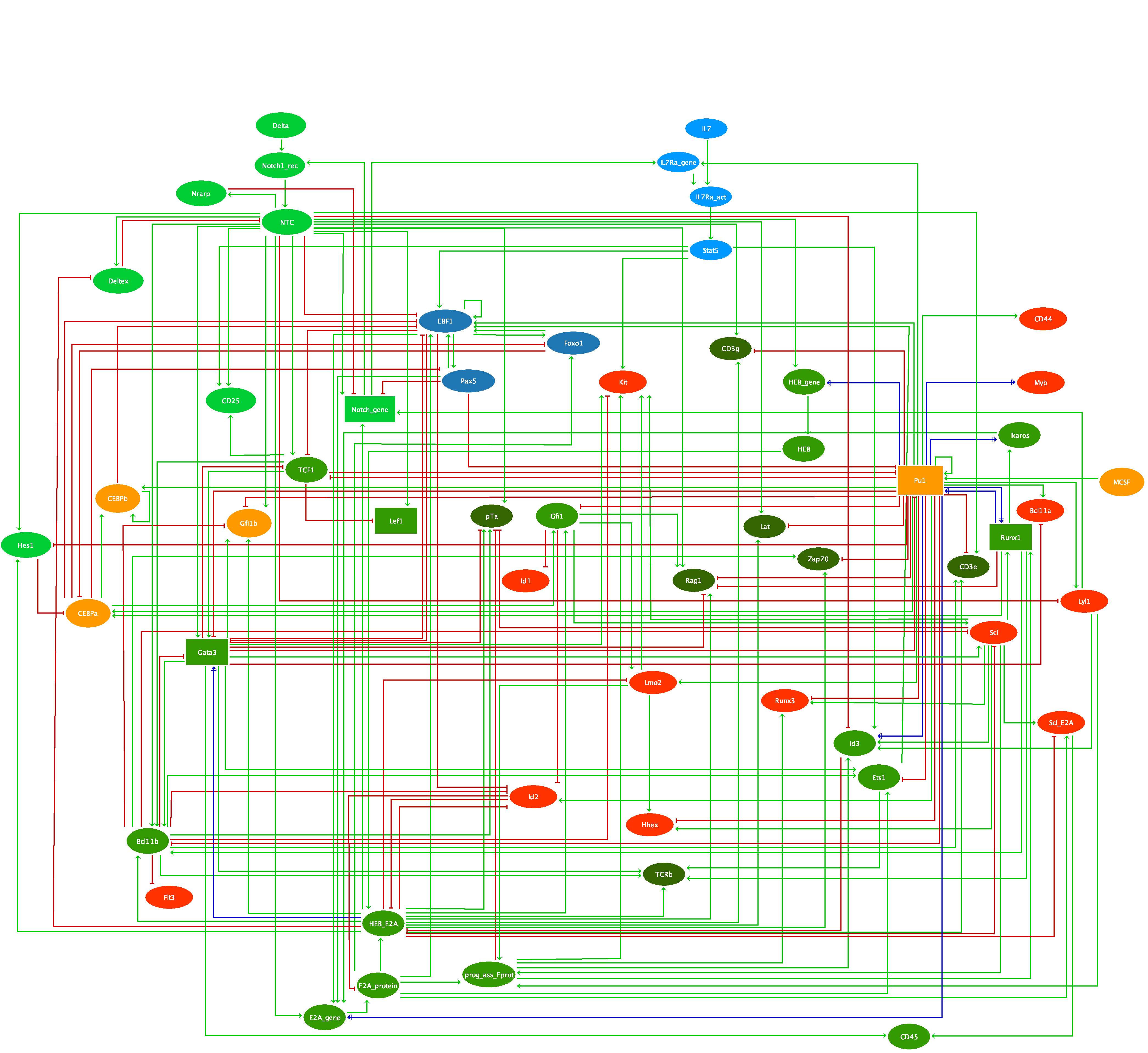
We have applied the logical modelling framework to the regulatory network controlling T-lymphocyte specification. This process involves cross-regulations between specific T-cell regulatory factors with factors driving alternative differentiation pathways, which remain accessible during the early steps of thymocyte development. Many transcription factors needed for T-cell specification are required in other hematopoietic differentiation pathways, and are combined in a fine-tuned, time-dependent fashion to achieve T-cell commitment.
Using the software GINsim, we integrated current knowledge into a dynamical model, which recapitulates the main developmental steps from early progenitors entering the thymus up to T-cell commitment, as well as the impact of various documented environmental and genetic perturbations. Our model analysis further enabled the identification of several knowledge gaps.

The associated notebook can be loaded using the CoLoMoTo notebook docker image (see http://www.colomoto.org/notebook).

Jupyter Notebook: Tdev_notebook_2nov2019.ipynb

Curation
Submitter: 
Pedro Monteiro

Drosophila mesoderm specification

Summary: 

This logical model encompasses 48 components and 82 regulatory interactions controlling mesoderm specification during Drosophila development, thereby integrating all major genetic processes underlying the formation of four mesodermal tissues. The model is based on in vivo genetic data, partly confirmed by functional genomic data.

Model simulations qualitatively recapitulate the expression of the main lineage markers of each mesodermal derivative, from developmental stage 8 to 10, for the wild type case, as well as for over twenty reported mutant genotypes.

This model has been used to systematically predict the effects of over 300 loss- and gain-off unction mutations, and combinations thereof. By generating specific mutant combinations, several novel predictions experimentally could be validated, demonstrating the robustness of model.

Curation
Submitter: 
Denis Thieffry

Molecular Pathways Enabling Tumour Cell Invasion and Migration

Summary: 

Understanding the etiology of metastasis is very important in clinical perspective, since it is estimated that metastasis accounts for 90% of cancer patient mortality. Metastasis results from a sequence of multiple steps including invasion and migration. The early stages of metastasis are tightly controlled in normal cells and can be drastically affected by malignant mutations; therefore, they might constitute the principal determinants of the overall metastatic rate even if the later stages take long to occur. To elucidate the role of individual mutations or their combinations affecting the metastatic development, a logical model has been constructed that recapitulates published experimental results of known gene perturbations on local invasion and migration processes, and predict the effect of not yet experimentally assessed mutations. The model has been validated using experimental data on transcriptome dynamics following TGF-β-dependent induction of Epithelial to Mesenchymal Transition in lung cancer cell lines. A method to associate gene expression profiles with different stable state solutions of the logical model has been developed for that purpose. In addition, we have systematically predicted alleviating (masking) and synergistic pairwise genetic interactions between the genes composing the model with respect to the probability of acquiring the metastatic phenotype. We focused on several unexpected synergistic genetic interactions leading to theoretically very high metastasis probability. Among them, the synergistic combination of Notch overexpression and p53 deletion shows one of the strongest effects, which is in agreement with a recent published experiment in a mouse model of gut cancer. The mathematical model can recapitulate experimental mutations in both cell line and mouse models. Furthermore, the model predicts new gene perturbations that affect the early steps of metastasis underlying potential intervention points for innovative therapeutic strategies in oncology.

Curation
Submitter: 
L. Calzone / C. Chaouiya

Mutually exclusive and co-occurring genetic alterations in bladder tumorigenesis

Summary: 

Relationships between genetic alterations, such as co-occurrence or mutual exclusivity, are often observed in cancer, where their understanding may provide new insights into etiology and clinical management. In this study, we combined statistical analyses and computational modelling to explain patterns of genetic alterations seen in 178 patients with bladder tumours (either muscle-invasive or non-muscle-invasive). A statistical analysis on frequently altered genes identified pair associations including co-occurrence or mutual exclusivity. Focusing on genetic alterations of protein-coding genes involved in growth factor receptor signalling, cell cycle and apoptosis entry, we complemented this analysis with a literature search to focus on nine pairs of genetic alterations of our dataset, with subsequent verification in three other datasets available publically. To understand the reasons and contexts of these patterns of associations while accounting for the dynamics of associated signalling pathways, we built a logical model. This model was validated first on published mutant mice data, then used to study patterns and to draw conclusions on counter-intuitive observations, allowing one to formulate predictions about conditions where combining genetic alterations benefits tumorigenesis. For example, while CDKN2A homozygous deletions occur in a context of FGFR3 activating mutations, our model suggests that additional PIK3CA mutation or p21CIP deletion would greatly favour invasiveness. Further, the model sheds light on the temporal orders of gene alterations, for example, showing how mutual exclusivity of FGFR3 and TP53 mutations is interpretable if FGFR3 is mutated first. Overall, our work shows how to predict combinations of the major gene alterations leading to invasiveness.

Curation
Submitter: 
Claudine Chaouiya

Mast cell activation

Summary: 

Based on an exhaustive curation of the existing literature and using the software CellDesigner, we have built and annotated a comprehensive molecular map for the FceRI and FcgRIIb signalling pathways, which play a key role in mast cell activation in mammals. Using this map and the logical modelling software GINsim, we have derived a logical model recapitulating the most salient features of mast cell activation. This model can be used to explore the dynamical properties of the system and its responses to different stimuli, in normal or mutant conditions. For more details, see [1].


References

Curation
Submitter: 
Denis Thieffry

MAPK network

Summary: 

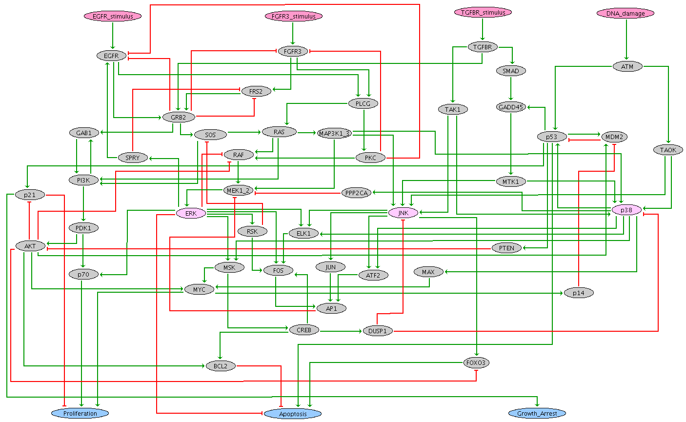
The Mitogen-Activated Protein Kinase (MAPK) network consists of tightly interconnected signalling pathways involved in the control of diverse cellular processes, including cell cycle, survival, apoptosis and differentiation.
Based on an extensive analysis of published data, we have built a comprehensive and generic reaction map for the MAPK signalling network, using CellDesigner software.

In order to explore the MAPK responses to different stimuli and better understand their contributions to cell fate decision, we have considered the most crucial components and interactions and encoded them into a logical model, using the software GINsim. Our logical model analysis particularly focuses on urinary bladder cancer, where MAPK network deregulations have often been associated with specific phenotypes.

To cope with the combinatorial explosion of the number of states, we have applied novel algorithms for model reduction and for the compression of state transition graphs, both implemented into the software GINsim. The results of systematic simulations for different signal combinations and network perturbations were found globally coherent with published data. In silico experiments further enabled us to delineate the roles of specific components, cross-talks and regulatory feedbacks in cell fate decision. Finally, tentative proliferative or anti-proliferative mechanisms can be connected with established bladder cancer deregulations, namely Epidermal Growth Factor Receptor (EGFR) over-expression and Fibroblast Growth Factor Receptor 3 (FGFR3) activating mutations.


Curation
Submitter: 
Denis Thieffry (P.T. Monteiro)

Drosophila VEGF Signalling pathway

Summary: 

VEGF (also called PDGF or PVF) pathway participates in different developmental processes, including border cell migration, hemocyte migration and survival, thorax closure during metamorphosis, the rotation and dorsal closure of the male terminalia. and embryonic salivary gland tissue migration. The ability of PVR to activate the MAP-kinase pathway is important for control of cell growth and differentiation in other tissues. Three genes in the Drosophila genome code for PVR ligands: PVF1, PVF2, and PVF3. Binding of one of the ligands (PVF1, 2 or 3) to the receptor PVR triggers the canonical DRK/SOS/RAS/RAF/DSOR1/RL pathway ([1];[2]; [3]; [4]; [5]). DOF is needed to assemble the PVR receptor and allow it to auto-phosphorylate, likely as an adaptor that links the receptor to RAS pathway. DOF is a cytoplasmic protein which is expressed ubiquitously only in cells that express the FGF receptors. It contains an ankyrin repeat, a coiled-coil structure and many tyrosines within environments that suggest that if phosphorylated they act as binding sites for the SH2 domains of proteins such as DRK or CSW ([6]). The SH2-domain-containing protein DRK recruits the guanine nucleotide exchange factor, Son of sevenless (SOS), to catalyze the exchange of GDP bound to RAS for GTP, thereby activating RAS with the help of activated KSR. RAS promotes the activation of RAF, leading to the activation of DSOR1, and ultimately to that of the MAP kinase Rolled (RL ). Rolled can activate transcription, both through inactivation of transcriptional co-repressors such as AOP, as well as through the activation of transcription factors such as the ETS-domain- containing protein Pointed (PNT) ([7]; [8]). The activation of PNT is a major output of the pathway. It is either phosphorylated by MAP kinase to produce an active transcriptional activator (PointedP2), or transcriptionally induced by MAP kinase to produce a constitutive transcriptional activator (PointedP1). Sprouty (STY) acts downstream of the receptor, but upstream of RAS1 and RAF, by recruiting GAP1 and blocking the ability of DRK to bind to its positive effector. We have considered three typical initial states corresponding to i. ligands binding in wild-type signalling enabling situation (VEGF_signalling), ii. ligand binding in the presence of the inhibitor Sprouty (Sprouty_inhibition), iii. absence of ligand (No_signalling).


References

Curation
Submitter: 
Abibatou MBODJ and Denis THIEFFRY

Drosophila Toll Signalling pathway

Summary: 

Toll was initially discovered as an essential component of the pathway that establishes the dorsal, ventral axis of the early Drosophila embryo. If any component in that genetic pathway is missing, no ventral or lateral cell types develop and the resulting embryos lack all mesoderm and the entire nervous system ([1]). Fungal and Gram-positive bacterial infections in Drosophila also stimulate the Toll pathway. Activation of Toll leads to recruitment of three cytoplasmic proteins, which are MYD88, Tube and Pelle, to form the signalling complex underneath the cell membrane ([2]). Subsequently, through interactions via death domains, assembly of the signalling complex containing MYD88, Tube and Pelle occurs ([2]; [3]). From this complex, signalling proceeds through the phosphorylation and degradation of the Drosophila IkB factor Cactus. In non signaling conditions, Cactus is bound to Dorsal or Dorsal-related immunity factor (DIF), inhibiting their activity and nuclear localization. Pelle is the only kinase reported for Cactus phosphorylation. After phosphorylation, nuclear translocation of Dorsal/DIF leads to activation of transcription of several sets of target genes. ([3]; [4]). To reproduce pathway signalling dynamics, we define two initial states corresponding to no signalling conditions (no ligand binding) and to signalling conditions (binding of SPZ to the receptor Toll).


References

  1. Anderson KV.  2000.  Toll signaling pathways in the innate immune response.. Current opinion in immunology. 12(1):13-9.
  2. Sun H, Towb P, Chiem DN, Foster BA, Wasserman SA.  2004.  Regulated assembly of the Toll signaling complex drives Drosophila dorsoventral patterning.. The EMBO journal. 23(1):100-10.
  3. Tanji T, Ip TY.  2005.  Regulators of the Toll and Imd pathways in the Drosophila innate immune response.. Trends in immunology. 26(4):193-8.
  4. Valanne S, Wang J-H, Rämet M.  2011.  The Drosophila Toll signaling pathway.. Journal of immunology (Baltimore, Md. : 1950). 186(2):649-56.
Curation
Submitter: 
Abibatou MBODJ and Denis THIEFFRY

Drosophila Hh Signalling pathway

Summary: 

Processing of HH ligand The precursor of HH is auto-catalytically cleaved to produce an N-terminal (HH-N) and a C- terminal (HH-C) fragments ([1]; [2]). A cholesterol moiety is covalently attached to the last amino acid of HH-N to create HH-Np, that is responsible for the biological activities of HH proteins ([1]; [2]; [3]). The N-terminal region of HH-Np is further modified by addition of palmitate that is essential for its signalling activity ([4]; [5]; [6]; [7]; [8]; [9]). We model these aspects by an AND rule (combining inputs from DLP, IHOG, Rasp, DISP, SHF, Lipophorin, BOI and DALLY) attached to the component representing the secreted HH molecule, denoted Hh in our model. HH Signalling Two integral membrane proteins are involved in HH signal reception: Patched and Smoothened. HH binding to its receptor Patched (PTC) relieves PTC-mediated repression of Smoothened (SMO), a serpentine-like membrane protein required for HH signalling ([10]; [11]). This allows SMO stabilisation, activation, and phosphorylation by Shaggy (SGG), and downstream signalling through the formation of a protein complex including the serine threonine kinase Fused (FU), the kinesin-like protein Costa (COS), and the protein Suppressor of Fused (SU(FU)), ultimately controlling the post-translational processing of the protein Cubitus interruptus (CI) ([12]). In the absence of HH, COS binds CI directly and sequesters it in the cytoplasm with the help of SUFU. The recruitment of different kinases (Casein kinase 1 alpha, Shaggy, Protein kinase A) then leads to the phosphorylation of CI and to its proteolysis by SLMB. The resulting truncated protein (CI_rep) is released and enters the nucleus, where it has a transcriptional repressing activity. Recent evidence further indicates that SMO is inhibited by TOW, which tentatively mediates the effect of PTC on SMO ([13]; [14]). Following SMO activation, the transcription factor CI is phosphorylated and translocated into the nucleus in its entire form, which plays a transcriptional activatory role (CI_act). In the model, a cascade of inhibitions, from HH on PTC, and from PTC on SMO, implements the indirect positive action of HH on SMO. A protein complex including CI, COS, and FU, phosphorylates and thereby inhibits SU(FU), ultimately favouring the CI activatory form and its translocation into the nucleus. We model the roles of the kinases (SGG, PKA, and CK1a), COS and SU(FU) (both needed to recruit the kinases) in the processing of CI in terms of inhibitory interactions on CI_act and activatory interactions on CI_rep ([15]; [16]). Complexes are represented implicitly (they are formed as soon as the components are synthesised or activated), while logical rules define component activity requirements to form CI_act versus CI_rep forms. To explore the dynamic of the pathway, we define two initial states to simulate the presence and the absence of signalling. On one hand, the non binding of HH (level expression 0) triggers a series of signalling cascades that lead to the activation of several kinases (for example SGG, PKA, CK1a, ...) at level of expression 1, which will permit the formation of CI repressor (expressed at level 1), which in turn will inhibit the targets. On the other hand, the presence of HH (level of expression 1) leads to a stable state corresponding to the signalling conditions leading to the formation of CI activator that will activate the targets node (level of expression 1).


References

  1. Lee JJ, Ekker SC, von Kessler DP, Porter JA, Sun BI, Beachy PA.  1994.  Autoproteolysis in hedgehog protein biogenesis.. Science (New York, N.Y.). 266(5190):1528-37.
  2. Porter JA, Young KE, Beachy PA.  1996.  Cholesterol modification of hedgehog signaling proteins in animal development.. Science (New York, N.Y.). 274(5285):255-9.
  3. Ingham PW, McMahon AP.  2001.  Hedgehog signaling in animal development: paradigms and principles.. Genes & development. 15(23):3059-87.
  4. Pepinsky RB, Zeng C, Wen D, Rayhorn P, Baker DP, Williams KP, Bixler SA, Ambrose CM, Garber EA, Miatkowski K et al..  1998.  Identification of a palmitic acid-modified form of human Sonic hedgehog.. The Journal of biological chemistry. 273(22):14037-45.
  5. Wang G, Amanai K, Wang B, Jiang J.  2000.  Interactions with Costal2 and suppressor of fused regulate nuclear translocation and activity of cubitus interruptus.. Genes & development. 14(22):2893-905.
  6. Amanai K, Jiang J.  2001.  Distinct roles of Central missing and Dispatched in sending the Hedgehog signal.. Development (Cambridge, England). 128(24):5119-27.
  7. Chamoun Z, Mann RK, Nellen D, von Kessler DP, Bellotto M, Beachy PA, Basler K.  2001.  Skinny hedgehog, an acyltransferase required for palmitoylation and activity of the hedgehog signal.. Science (New York, N.Y.). 293(5537):2080-4.
  8. Lee JD, Treisman JE.  2001.  The role of Wingless signaling in establishing the anteroposterior and dorsoventral axes of the eye disc.. Development (Cambridge, England). 128(9):1519-29.
  9. Micchelli CA, The I, Selva E, Mogila V, Perrimon N.  2002.  Rasp, a putative transmembrane acyltransferase, is required for Hedgehog signaling.. Development (Cambridge, England). 129(4):843-51.
  10. Alcedo J, Ayzenzon M, Von Ohlen T, Noll M, Hooper JE.  1996.  The Drosophila smoothened gene encodes a seven-pass membrane protein, a putative receptor for the hedgehog signal.. Cell. 86(2):221-32.
  11. Chen Y, Struhl G.  1996.  Dual roles for patched in sequestering and transducing Hedgehog.. Cell. 87(3):553-63.
  12. Lum L, Yao S, Mozer B, Rovescalli A, Von Kessler D, Nirenberg M, Beachy PA.  2003.  Identification of Hedgehog pathway components by RNAi in Drosophila cultured cells.. Science (New York, N.Y.). 299(5615):2039-45.
  13. Ayers KL, Rodriguez R, Gallet A, Ruel L, Thérond P.  2009.  Tow (Target of Wingless), a novel repressor of the Hedgehog pathway in Drosophila.. Developmental biology. 329(2):280-93.
  14. Ayers KL, Thérond PP.  2010.  Evaluating Smoothened as a G-protein-coupled receptor for Hedgehog signalling.. Trends in cell biology. 20(5):287-98.
  15. Aikin RA, Ayers KL, Thérond PP.  2008.  The role of kinases in the Hedgehog signalling pathway.. EMBO reports. 9(4):330-6.
  16. Wilson CW, Chuang P-T.  2006.  New "hogs" in Hedgehog transport and signal reception.. Cell. 125(3):435-8.
Curation
Submitter: 
Abibatou MBODJ and Denis THIEFFRY

Drosophila SPATZLE Processing pathway

Summary: 

During DV patterning, a regulatory cascades composed by three dorsal group genes gastrulation-defective, snake and easter, encoding serine proteases, lead to the cleavage of Spatzle (SPZ), that in turn activates the Toll-dorsal signaling pathway ([1]; [2]). Spatzle presumably forms a gradient in the perivitelline fluid. Toll signaling is ultimately responsible for the formation of the embryonic dorsal nuclear gradient. In the nucleus, dorsal controls the expression of zygotic genes in a concentration-dependent manner and this process results in the patterning of the dorsal–ventral embryonic axis. twist is one of the earliest target genes controlled by the highest concentration of dorsal in the mesodermal cells. It is a transcriptional activator that cooperates with dorsal in activating snail in the mesoderm. Dorsal and Twist also cooperate to activate the neurogenic gene, sim (single minded), expressed in the neurectoderm and repressed by Snail in the mesoderm. Natural or experimentally induced infections by fungi or bacteria elicit a specific response in both adult flies and larvae. The proteoglycans of Gram-positive and Gram-negative bacteria are sensed by distinct pattern recognition proteins called PGRPs (peptidoglycan recognition proteins ([3]). Different PRGPs cooperate to activate the Toll pathway. The activation of PGRP-SA by Gram- positive bacteria leads to Spatzle cleavage ([4]). Fungal infection also leads to the cleavage of Spatzle, but the proteolytic cascade in this case involves the circulating serine protease Persephone and its serine protease inhibitor, Necrotic ([5]; [6]; [7]). Circulating PGRP-SA receptor activates the Toll pathway upon detection of Lysine-type PGN which is a major component of the cell wall of many Gram-positive bacterial strains. GNBP1 (Gram-Negative Binding Protein 1) associates with PGRP-SA and this complex activates a downstream proteolytic cascade that leads to the cleavage of Spatzle, which then activates the Toll transmembrane receptor. In addition, four other serine proteases, namely Spirit, Spheroide, and Sphinx1 and 2, were identified in response to both fungi and Gram-positive bacteria infections. Thus, PGRP-SA and GNBP1 define a Gram-positive-specific branch of Toll receptor activation. PGRP-SD also belongs to this branch and is required for the detection of other Gram-positive and negative bacterial strains. In short, the maturation of SPZ activates Toll in both early embryo and immune response and is controlled by different sets of proteases ([8]; [9]). To reproduce biological data during SPZ processing, we define four initial states corresponding the biological process involved. All these initial state lead to the formation of the active form of SPZ.


References

  1. Morisato D, Anderson KV.  1994.  The spätzle gene encodes a component of the extracellular signaling pathway establishing the dorsal-ventral pattern of the Drosophila embryo.. Cell. 76(4):677-88.
  2. Weber ANR, Tauszig-Delamasure S, Hoffmann JA, Lelièvre E, Gascan H, Ray KP, Morse MA, Imler J-L, Gay NJ.  2003.  Binding of the Drosophila cytokine Spätzle to Toll is direct and establishes signaling.. Nature immunology. 4(8):794-800.
  3. Royet J.  2004.  Drosophila melanogaster innate immunity: an emerging role for peptidoglycan recognition proteins in bacteria detection.. Cellular and molecular life sciences : CMLS. 61(5):537-46.
  4. Gobert V, Gottar M, Matskevich AA, Rutschmann S, Royet J, Belvin M, Hoffmann JA, Ferrandon D.  2003.  Dual activation of the Drosophila toll pathway by two pattern recognition receptors.. Science (New York, N.Y.). 302(5653):2126-30.
  5. Ligoxygakis P, Pelte N, Ji C, Leclerc V, Duvic B, Belvin M, Jiang H, Hoffmann JA, Reichhart J-M.  2002.  A serpin mutant links Toll activation to melanization in the host defence of Drosophila.. The EMBO journal. 21(23):6330-7.
  6. Ligoxygakis P, Pelte N, Hoffmann JA, Reichhart J-M.  2002.  Activation of Drosophila Toll during fungal infection by a blood serine protease.. Science (New York, N.Y.). 297(5578):114-6.
  7. Pelte N, Robertson AS, Zou Z, Belorgey D, Dafforn TR, Jiang H, Lomas D, Reichhart J-M, Gubb D.  2006.  Immune challenge induces N-terminal cleavage of the Drosophila serpin Necrotic.. Insect biochemistry and molecular biology. 36(1):37-46.
  8. Bischoff V, Vignal C, Boneca IG, Michel T, Hoffmann JA, Royet J.  2004.  Function of the drosophila pattern-recognition receptor PGRP-SD in the detection of Gram-positive bacteria.. Nature immunology. 5(11):1175-80.
  9. Valanne S, Wang J-H, Rämet M.  2011.  The Drosophila Toll signaling pathway.. Journal of immunology (Baltimore, Md. : 1950). 186(2):649-56.
Curation
Submitter: 
Abibatou MBODJ and Denis THIEFFRY
Syndicate content


about seo